This study was conducted with Dong Hun Lee and the co-corresponding authors listed below.
- Yong-Hee Kim
- Department of Basic Research, PB Immune Therapeutics Inc, Seoul, Republic of Korea
- Transplantation Research Institute, Seoul National University Medical Research Center, Seoul, Republic of Korea
- Hyun Je Kim
- Department of Biomedical Sciences, Seoul National University College of Medicine, Seoul, Republic of Korea
- Department of Microbiology and Immunology, Seoul National University College of Medicine, Seoul, Republic of Korea
- Cancer Research Institute, Seoul National University College of Medicine, Seoul, Republic of Korea
- Interdisciplinary Program in Artificial Intelligence (IPAI), Seoul National University, Seoul, Republic of Korea
- Genomic Medicine Institute, Medical Research Center, Seoul National University College of Medicine, Seoul, Republic of Korea
- Department of Dermatology, Seoul National University Hospital, Seoul National University College of Medicine, Seoul, Republic of Korea
- Dong Hun Lee / EASI Lab PI
- Institute of Human-Environment Interface Biology, Medical Research Center, Seoul National University, Seoul, Republic of Korea
- Department of Dermatology, Seoul National University Hospital, Seoul National University College of Medicine, Seoul, Republic of Korea
Names and affiliations are as published in the article.
The question
Do expanded regulatory T cells in atopic dermatitis retain effective suppression, or do they contain distinct functional states?
How we tested it
PBMCs from 48 people with AD and 48 controls were profiled using 39-parameter CyTOF and clustering. Reanalysis of public PBMC and skin scRNA-seq was combined with in vitro OX40⁺ Treg suppression assays and tissue immunofluorescence.
CyTOF, functional assays and skin immunofluorescence were performed in this collaborative study; scRNA-seq evidence came from public-data reanalysis. Functional assays involved 5 HC and 3 AD donors, and skin immunofluorescence involved 7 participants per group.
What the study found
A Treg population with high OX40 and CRTH2 expression was associated with disease severity. OX40⁺ Tregs from AD donors showed lower suppressive capacity than the corresponding OX40⁺ population from controls, with enrichment also supported in skin.
Interpreting the evidence
The main cohort was Korean and the functional assays involved few donors. In vitro findings do not fully reproduce the skin microenvironment, and the study does not establish prediction of responses to OX40-targeted treatment.
Figures from the paper
Complete Figure 6a–f: complementary evidence from public skin scRNA-seq and tissue immunofluorescence. CyTOF and suppression assays are not shown in this figure.
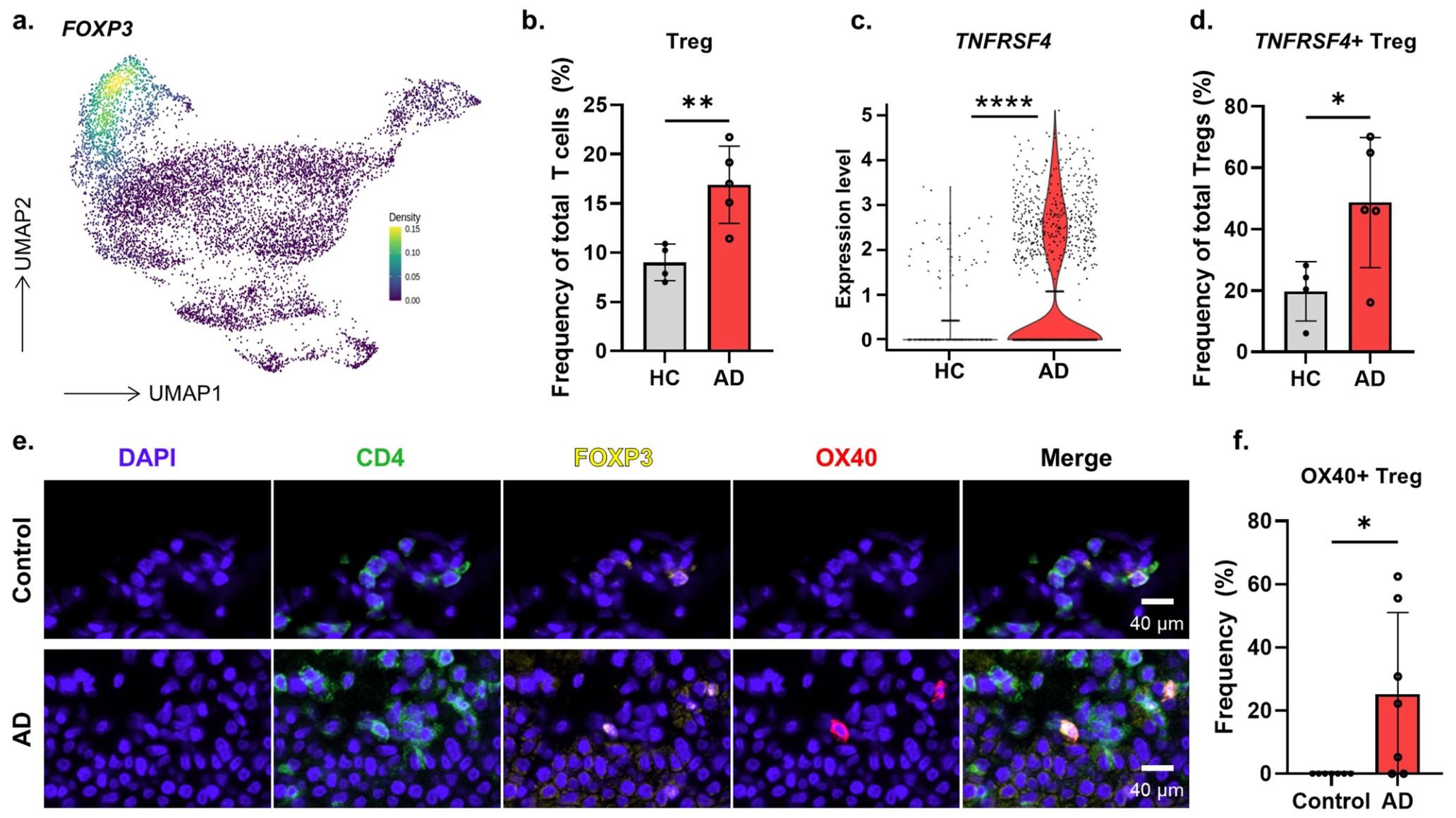
Public skin single-cell data and tissue immunofluorescence provide complementary evidence for OX40⁺ regulatory T cells in atopic dermatitis. Shown: the complete original Figure 6.
© 2026 Japanese Society of Allergology. Published by Elsevier B.V. CC BY-NC-ND 4.0. CC BY-NC-ND 4.0 · PublicationA question to explore together
Could longitudinal sampling of skin and blood reveal how changes in OX40⁺ Treg states relate to clinical response?
Discuss this question Explore collaboration topics